Treatment Brief
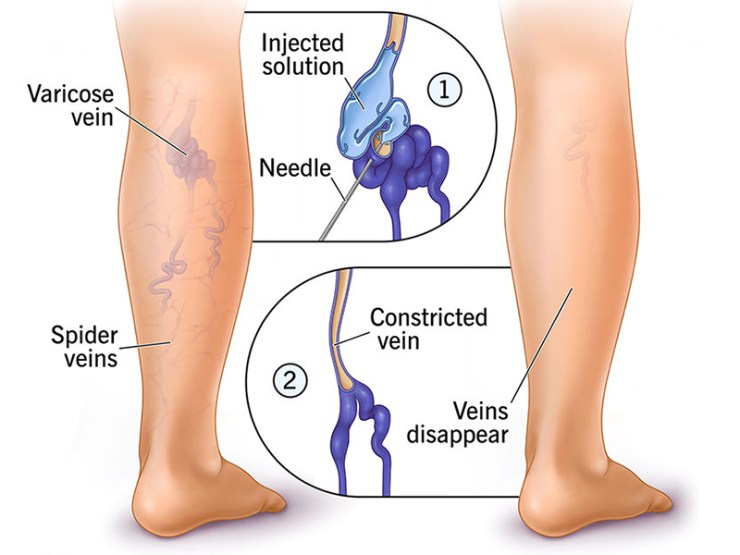
What is foam sclerotherapy?
If we’re treating large varicose veins, we’ll use ultrasound guidance during your procedure. This isn’t usually necessary for smaller veins that are close to the surface, as visual guidance is sufficient.
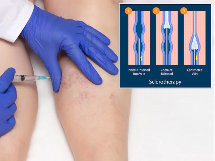
Due to the series of tiny injections involved in the foam sclerotherapy procedure, some patients find them uncomfortable or mildly painful. The procedure usually takes around 30-45 minutes.
Post-Procedure
Following your treatment, your nurse will help you put on a compression stocking, which you need to wear for 7 days after your procedure. You’ll wear two of these if you have both legs treated. Your nurse will explain how to wear these and bathe in them. They’ll also provide you with aftercare advice, including any post-treatment symptoms to be aware of.
You’re encouraged to go for a walk for at least 10 minutes prior to returning home.
What happens after treatment?
You should keep the compression pads, bandage, and stocking on continuously for 5 days. After this you may remove the pads and bandage and then replace the stocking which should be worn for a further 7 days. During this 7 day period, you may remove the stocking to shower and you may remove it at night if you wish. If you find the stocking comfortable and wish to wear it for longer, this may be helpful. Please bring your stocking back with you to your next visit as it may be possible to reuse it if you have further injections.
You should do plenty of walking and may generally do most normal activities without any problem. If in doubt ask your doctor.
You can be active as usual after the treatment and do not need to avoid anything in particular. However you should not drive on the day the procedure is performed, just in case you experience any visual disturbances (see below).
Flow

Advantage
Compatible with Imaging
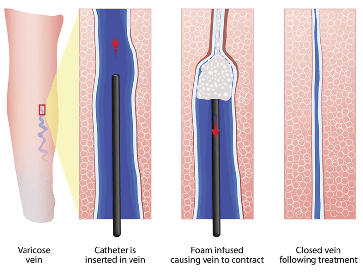
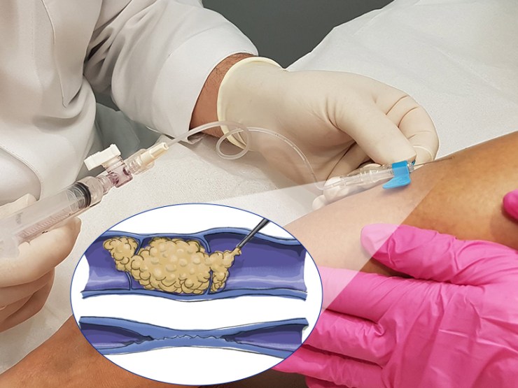
Another unique benefit of foam sclerotherapy is the physical makeup of the foam sclerant. The difference in density when compared to blood allows for the sclerant to be detected using different types of imaging.
This can help our better evaluate how effective the sclerant is in facilitating a clot. Ultrasonic imaging can help determine how effective the sclerant is. This can expedite the process of treating your veins if additional treatments are needed.


